Íomhá:Schematic of cortical areas involved with pain processing and fMRI cropped.jpg

Size of this preview: 800 × 541 picteilín. Other resolutions: 320 × 216 picteilín | 640 × 432 picteilín | 1,024 × 692 picteilín | 1,172 × 792 picteilín.
Taispeáin leagan ardtaifigh den íomhá (1,172 × 792 picteilín, méid comhaid: 211 KB, cineál MIME: image/jpeg)
| Seo comhad as An Cómhaoin Viciméid. Tá an tuairisc as an leathanach tuairisc Cómhaoin a leanas thíos.
|
| Tuairisc |
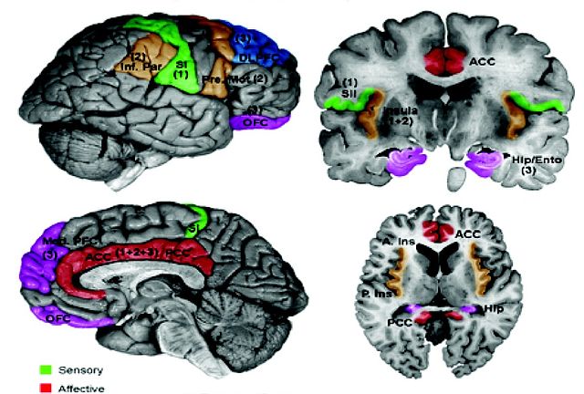
English: Examples of CNS Functional Measures. A. Schematic of cortical areas involved with pain processing. The highlighted areas summarize areas found active in previous functional imaging studies. Color-coding reflects the hypothesized role of each area in processing the different psychological dimensions of pain. Numbers in parentheses indicate the relative involvement of these areas during different temporal stages of the pain experience. Areas displayed include insula, anterior cingulate cortex (ACC), posterior cingulate cortex (PCC), primary somatosensory cortex (SI), secondary somatosensory cortex (SII), inferior parietal lobe (Inf. Par), dorsolateral prefrontal cortex (DLPFC), pre-motor cortex (Pre-Mot), orbitofrontal cortex (OFC), medial prefrontal cortex (Med. PFC), posterior insula (P. Ins), anterior insula (A. Ins), hippocampus (Hip), entorhinal cortex (Ento). [Reprinted with permission from Casey and Tran, 2006]. For examples of brainstem involvement in pain processing, please refer to Tracey and Iannetti ([52]). B. Example of fMRI responses to painful phasic thermal stimulation to the forehead in a cohort of 12 subjects. (Moulton et al., unpublished observations). Borsook et al. Molecular Pain 2007 3:25 doi:10.1186/1744-8069-3-25 |
| Dáta | (UTC) |
| Foinse | |
| Údar |
|
| This is a retouched picture, which means that it has been digitally altered from its original version. Modifications: Cropped. The original can be viewed here: Schematic of cortical areas involved with pain processing and fMRI.jpg:
|
Is mise sealbhóir cóipchirt an shaothair seo agus foilsím é faoin gceadúnas seo a leanas:
This file is licensed under the Creative Commons Attribution 2.0 Generic license.
- Is féidir:
- an saothar seo a roinnt – an saothar seo a chóipeáil, a dháil amach, agus a tharchur
- an saothar seo a athmheascadh – an saothar seo a oiriúnú
- Ach amháin má leantar na coinníollacha seo a leanas:
- sannadh – Ní mor creidiúint a thabhairt leis an saothar, faoi mar a shonraigh an údar nó tabharthóir an ceadúnais (ach ná húsáid modh a thugann le fios go thacaíonn sé/sí tú féin nó an úsáid a bhaineann tú as an saothar).
Original upload log
This image is a derivative work of the following images:
- File:Schematic_of_cortical_areas_involved_with_pain_processing_and_fMRI.jpg licensed with Cc-by-2.0
- 2009-08-27T15:01:26Z CopperKettle 1200x1383 (386847 Bytes) {{Information |Description={{en|1=Examples of CNS Functional Measures. A. Schematic of cortical areas involved with pain processing. The highlighted areas summarize areas found active in previous functional imaging studies. C
Uploaded with derivativeFX
Stair chomhaid
Cliceáil ar dáta/am chun an comhad a radharc mar a bhí sé ag an am sin.
| Dáta/Am | Mionsamhail | Toisí | Úsáideoir | Nóta tráchta | |
|---|---|---|---|---|---|
| faoi láthair | 13:39, 5 Meitheamh 2010 | | 1,172 × 792 (211 KB) | Anthonyhcole | {{Information |Description={{en|1=Examples of CNS Functional Measures. A. Schematic of cortical areas involved with pain processing. The highlighted areas summarize areas found active in previous functional imaging studies. Color-coding reflects the hypot |
Úsáid comhaid
Tá nasc chuig an gcomhad seo ar na leathanaigh seo a leanas:
Global file usage
The following other wikis use this file:
- Úsáid ar en.wikipedia.org
- Úsáid ar en.wikiquote.org
- Úsáid ar en.wikiversity.org
- Úsáid ar es.wikipedia.org
- Úsáid ar it.wikipedia.org
- Úsáid ar ru.wikiversity.org